Critical Cardiac Emergencies
A Structured Approach to High-Risk Cardiovascular Presentations | 2026
Educational resource for SASEM training. Not a substitute for clinical judgment, current AHA/ESC/ACC guidelines, or hospital protocols.
ABDULRAHMAN ALZAHRANI, MD, SBEM
Emergency Medicine Consultant
Important Notice
This presentation is for education only and does not replace clinical judgment, local protocols, or official guidelines. Use patient-specific assessment and institutional policies for care decisions.
Start Here
Quiz 16Q
Quick pre-quiz check
Assessment
Password-protected assessment
MCQ 5
Advanced questions with explanations
CPR Helper
Timer & Metronome
Cases
Step-by-step interactive cases
Calculators
HEART, Wells, CHA₂DS₂
The Cardiac Emergency Spectrum
Categorizing Life Threats Beyond Simple Chest Pain
Core Categories of Cardiovascular Collapse
Ischemia
ACS, Myocardial Injury
Electrical
Arrest, Tachy, Brady
Pump Failure
Heart Failure, Cardiogenic Shock
Mechanical
Tamponade, Valve Rupture
Valvular
Acute Valve Failure
Vascular
Dissection, AAA, PE
Pressure
Hypertensive Crisis
Syncope
Cardiogenic Causes
Summary
Key Takeaways & Review
Initial Approach
Stabilize, Assess, Triage
The First 10 Minutes & Beyond
Priority Actions
- ABC's & Stability: Secure airway, ensure adequate breathing, and assess for shock (pulse, perfusion).
- IV Access & Monitor: Two large-bore IVs, place on continuous cardiac monitor and pulse oximetry.
- ECG within 10 minutes: Essential for all high-risk presentations (Chest Pain, Syncope, Dyspnea).
Focused Workup
- POCUS: Rapidly assess for tamponade, severe RV strain, or LV dysfunction.
- Labs: Troponin, BNP, D-dimer, Electrolytes, Hgb.
- Serial Reassessment: Symptoms and ECGs are dynamic. Repeat evaluation is key.
ACS 1: Introduction to Acute Coronary Syndromes
Understanding Myocardial Ischemia Spectrum
🔍 What is an Acute Coronary Syndrome?
Acute Coronary Syndrome (ACS) is a spectrum of conditions compatible with acute myocardial ischemia and/or infarction, usually due to coronary artery disease.
📊 ACS Epidemiology
Global Burden
- 8.9 million deaths annually worldwide
- Leading cause of death globally
- 30% of all deaths in adults >35 years
- Increasing incidence in young adults
Time is Muscle
- Each hour delay = 1% mortality increase
- Door-to-balloon goal: ≤90 minutes
- First medical contact: ≤10 minutes
- Early recognition saves lives
🔬 Pathophysiology of Myocardial Ischemia
The Ischemic Cascade
⚡ Clinical Presentation
Typical Chest Pain
- Retrosternal pressure/discomfort
- Radiates to left arm, jaw, neck
- Duration >20 minutes
- Not relieved by rest or NTG
- Associated with diaphoresis, nausea, dyspnea
Atypical Presentations
- Women: Epigastric pain, fatigue, nausea
- Diabetics: Silent ischemia, atypical symptoms
- Elderly: Weakness, confusion, syncope
- Post-op: Incisional pain masking cardiac pain
Key Learning Points
- ACS is a spectrum: STEMI → NSTEMI → Unstable Angina (increasing severity)
- Time-critical diagnosis: ECG within 10 minutes of presentation
- Chest pain ≠ cardiac pain: Consider differential diagnosis
- High clinical suspicion: Better to over-treat than miss ACS
- Patient factors matter: Age, gender, comorbidities affect presentation
ACS 2: STEMI ECG Patterns
Interactive Guide to Coronary Territories
| MI Type | Cause | Description |
|---|---|---|
| Type 1 | Atherosclerotic Plaque Rupture | Spontaneous MI due to primary coronary artery disease. |
| Type 2 | Supply-Demand Imbalance | MI due to increased oxygen demand or decreased supply (e.g., hypotension, anemia, arrhythmias). |
| Type 3 | MI leading to Death | Cardiac arrest or death before troponin samples are available. |
| Type 4 | PCI Related | MI occurring during or shortly after percutaneous coronary intervention. |
| Type 5 | CABG Related | MI occurring during or shortly after coronary artery bypass grafting. |
STEMI Criteria (Key Takeaways)
New ST-elevation in ≥ 2 contiguous leads. Contemporary guidelines recommend: ≥ 1.5 mm in V2-V3 (anterior leads); ≥ 1 mm in other leads. Gender-specific thresholds are no longer recommended as they may delay treatment.
STEMI Localization & ECG Patterns
Click on each territory to see ECG characteristics and clinical pearls.
Anterior STEMI
LAD Artery (V1-V4)
ST ≥1.5mm in V2-V3, ≥1mm in V1,V4-V6
Highest mortality - proximal LAD occlusion
Lateral STEMI
Circumflex Artery (I, aVL, V5-V6)
ST ≥1mm in lateral leads
Often missed - check lateral leads!
Inferior STEMI
RCA (85%) or LCx (15%)
ST ≥1mm in II, III, aVF
Check V4R for RV involvement
Posterior STEMI
Posterior Descending (V7-V9)
ST depression V1-V3 + tall R waves
Recommendation: Obtain V7-V9 leads (STE ≥0.5mm)
STEMI ECG Gallery
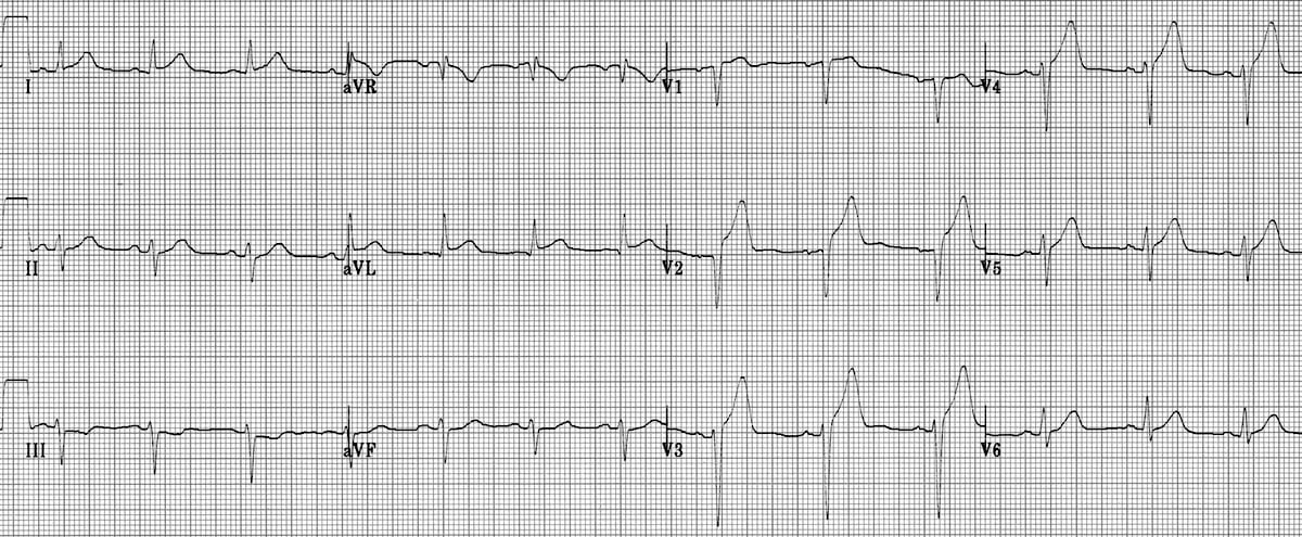
Anterior STEMI - ST elevation V1-V4
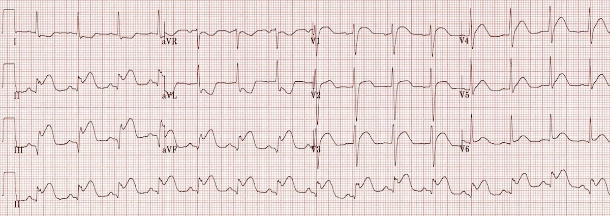
Inferior STEMI - ST elevation II, III, aVF
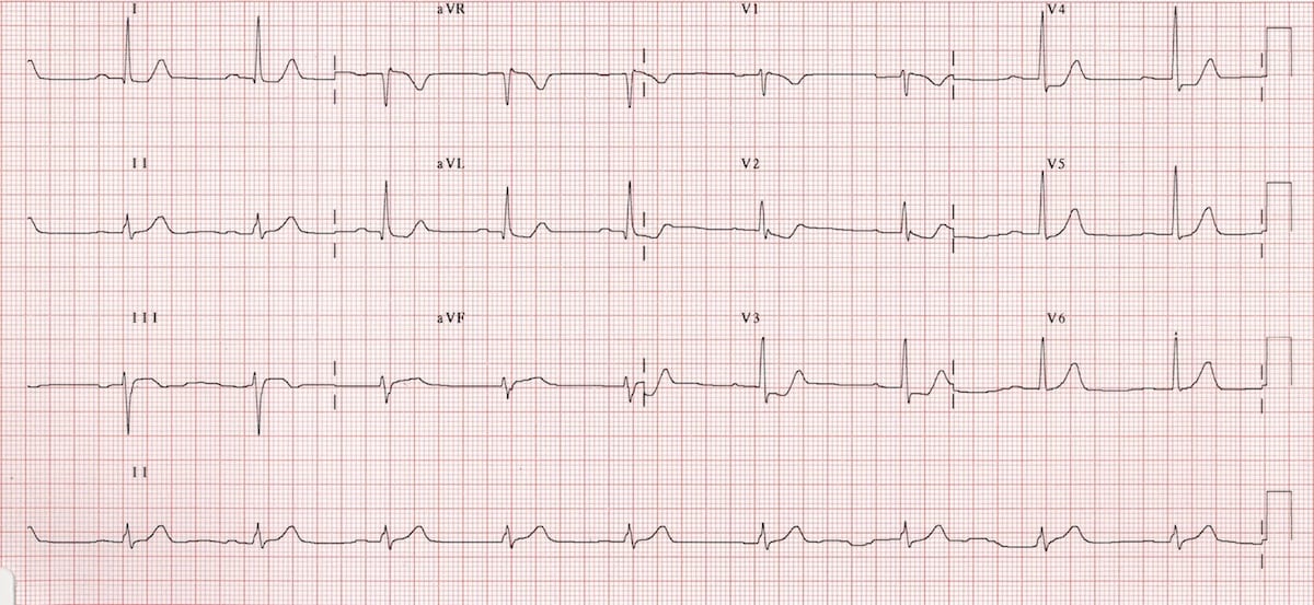
Posterior STEMI - ST depression V1-V3 + tall R waves
Critical Reminders
- Women & Diabetics: May have atypical presentations
- Right-sided ECG: For inferior STEMI with hypotension
- Posterior leads: V7-V9 if posterior STEMI suspected
- ST criteria: ≥1mm in most leads, ≥1.5mm in V2-V3
ACS 3: NSTEMI & Unstable Angina ECG Patterns
Non-ST-Elevation Myocardial Ischemia
🔍 NSTEMI vs Unstable Angina
NSTEMI (Non-ST-Elevation MI)
- Myocardial necrosis with elevated troponins
- ECG changes: ST depression, T-wave inversion
- Partial coronary occlusion
- Risk of progression to STEMI
- Requires urgent intervention
Unstable Angina
- Ischemia without necrosis (normal troponins)
- ECG changes: May be normal or nonspecific
- Coronary plaque disruption
- High risk of progression
- Requires risk stratification
📊 ECG Changes in NSTEMI
Common ECG Findings
ST Depression
- • ≥1mm in ≥2 contiguous leads
- • Often in leads I, aVL, V5-V6 (lateral)
- • May be reciprocal to ST elevation elsewhere
- • More common in inferior NSTEMI
T-Wave Inversion
- • ≥1mm deep in ≥2 contiguous leads
- • Often precedes ST depression
- • Most common finding in NSTEMI
- • May persist after resolution
🖼️ NSTEMI ECG Gallery
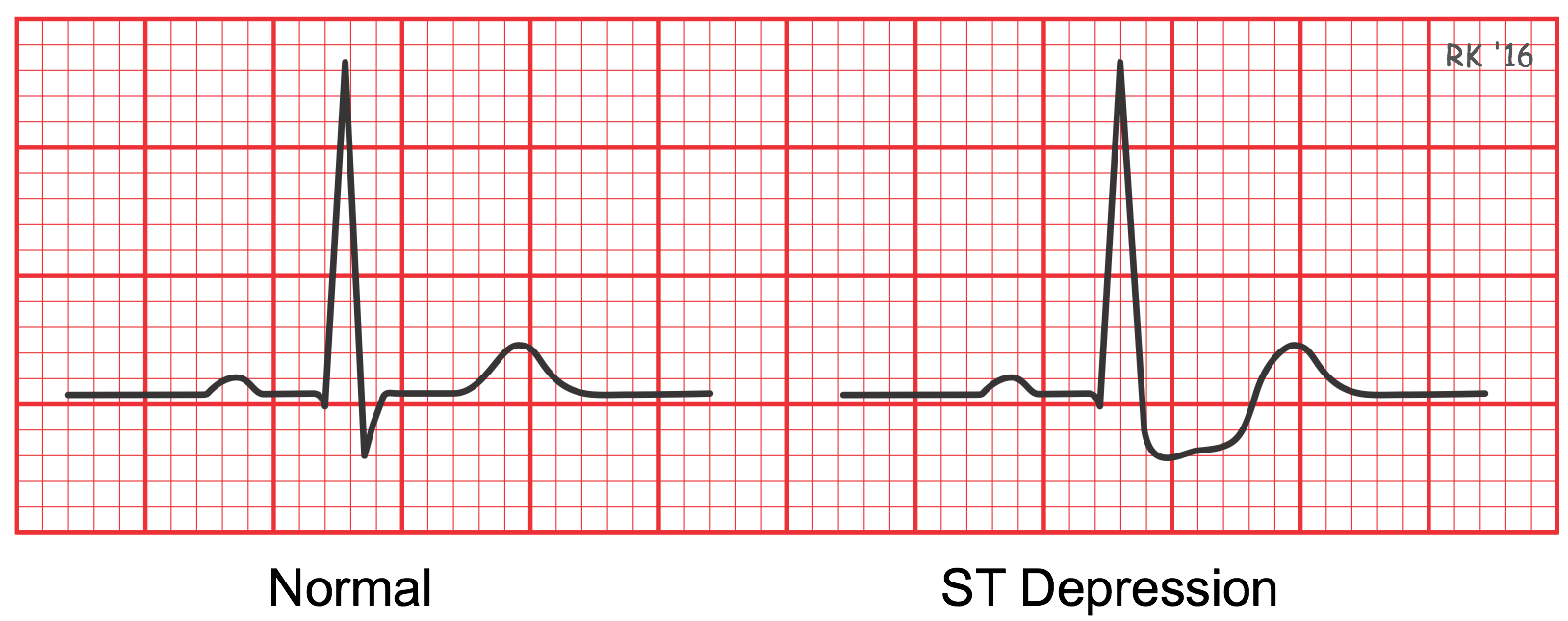
NSTEMI - ST depression in lateral leads
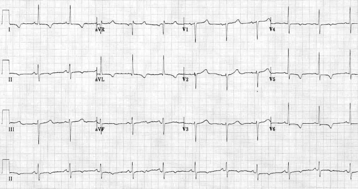
NSTEMI - T-wave inversion
⚠️ High-Risk ECG Features
Urgent Cath Lab Activation
- • ST depression ≥2mm in multiple leads
- • ST depression in anterior leads (V1-V4)
- • Dynamic ECG changes (new vs old)
- • ST depression with positive troponins
- • Modified Sgarbossa criteria in LBBB
- • Posterior ECG changes (tall R waves)
- • ECG changes in patients with prior CABG
- • Persistent symptoms despite medical therapy
Clinical Pearls
- Normal ECG doesn't rule out ACS: Up to 20% of NSTEMI have normal ECG initially
- Serial ECGs are crucial: Compare with old ECGs when available
- ST depression ≠ STEMI: But may indicate severe ischemia requiring urgent cath
- T-wave inversions evolve: May appear before ST depression in evolving ischemia
- Right-sided ECG: Consider for inferior NSTEMI with hypotension
ACS 4: Biomarkers in ACS
Troponins, CK-MB, and Risk Stratification
🧪 Cardiac Biomarkers Overview
Troponin I/T
Gold standard for myocardial injury
CK-MB
Historical marker, less specific
Myoglobin
Early marker, low specificity
🔬 High Sensitive Troponin (hs-Tn) - Worldwide Standard
Why High Sensitive Troponin Matters
🌍 Global Recommendation
- ESC 2023 ACS Guidelines: hs-cTn preferred for diagnosis and 0/1h–0/2h algorithms
- AHA/ACC 2021 Chest Pain: hs-cTn for early rule-out/rule-in pathways
- Universal adoption: Most labs worldwide use hs-Tn
⚡ Clinical Advantages
- Earlier detection: Detects injury 1-2 hours after onset
- Higher sensitivity: 10-100x more sensitive than conventional
- Better risk stratification: Detects minor myocardial injury
- Faster discharge: Enables 1-2 hour protocols
📊 hs-Tn Reference Ranges & Interpretation
| hs-Tn Level | Interpretation | Clinical Action |
|---|---|---|
|
Normal (< URL) |
No myocardial injury detected | Low suspicion: Consider discharge with outpatient follow-up |
|
Elevated (> URL but < 52 ng/L) |
Possible myocardial injury or chronic elevation | Serial testing required, assess delta change |
|
Rising
Pattern (>20% delta increase) |
Acute myocardial injury likely | Treat as NSTEMI, cardiology consultation |
|
Markedly
Elevated (>52 ng/L) |
Significant myocardial injury | Urgent cardiology evaluation, possible STEMI equivalent |
Key Points for hs-Tn Use
- Gender-specific cutoffs: Women have lower normal ranges than men
- Serial testing: Compare to baseline when available; look for rising trend
- Delta change: >20% increase indicates acute injury
- 1-2 hour protocols: Normal hs-Tn at 0h + 1-2h = rule-out MI
- Interference: Hemolysis, biotin supplements can falsely elevate
- Chronic elevations: CKD, HF patients may have baseline elevations
📈 Troponin Kinetics
| Biomarker | Onset | Peak | Return to Normal | Clinical Use |
|---|---|---|---|---|
| Troponin I | 3-6 hours | 12-24 hours | 5-10 days | Diagnosis of MI |
| Troponin T | 3-6 hours | 12-24 hours | 10-14 days | Diagnosis of MI |
| CK-MB | 3-6 hours | 12-24 hours | 2-3 days | Historical use |
| Myoglobin | 1-2 hours | 6-12 hours | 24 hours | Early detection |
🔬 Pathophysiology of Myocardial Ischemia
The Ischemic Cascade
⚡ Clinical Presentation
Typical Chest Pain
- Retrosternal pressure/discomfort
- Radiates to left arm, jaw, neck
- Duration >20 minutes
- Not relieved by rest or NTG
- Associated with diaphoresis, nausea, dyspnea
Atypical Presentations
- Women: Epigastric pain, fatigue, nausea
- Diabetics: Silent ischemia, atypical symptoms
- Elderly: Weakness, confusion, syncope
- Post-op: Incisional pain masking cardiac pain
Key Learning Points
- ACS is a spectrum: STEMI → NSTEMI → Unstable Angina (increasing severity)
- Time-critical diagnosis: ECG within 10 minutes of presentation
- Chest pain ≠ cardiac pain: Consider differential diagnosis
- High clinical suspicion: Better to over-treat than miss ACS
- Patient factors matter: Age, gender, comorbidities affect presentation
ACS 5: Risk Stratification in ACS
TIMI, GRACE, HEART Scores & Decision Making
📊 Risk Stratification Scores
TIMI Risk Score for NSTEMI/UA
1 point each for:
- • Age ≥65 years
- • ≥3 CAD risk factors
- • Known CAD (stenosis ≥50%)
- • ST deviation ≥0.5mm
- • ≥2 anginal episodes in 24h
- • ASA use in past 7 days
- • Elevated cardiac markers
HEART Score
Points (0-2 each):
- History: Highly suspicious (2), Moderately (1), Slightly (0)
- ECG: Significant ST depression (2), Nonspecific (1), Normal (0)
- Age: ≥65 (2), 45-64 (1), ≤44 (0)
- Risk factors: ≥3 (2), 1-2 (1), None (0)
- Troponin: ≥3x normal (2), 1-3x (1), Normal (0)
⚡ Management Based on Risk
Treatment Algorithms
High-Risk Features (Urgent Cath)
- • TIMI ≥4 or HEART ≥7
- • Recurrent ischemia at rest
- • ST depression ≥1mm
- • New/worsening MR
- • Elevated troponins
- • Hemodynamic instability
- • Sustained VT
Low-Risk Features (Medical Management)
- • TIMI 0-2 or HEART 0-3
- • No recurrent symptoms
- • Normal ECG
- • Normal troponins
- • No hemodynamic issues
- • Good functional capacity
💊 Anti-Ischemic Therapy
Initial Medical Therapy
- Aspirin 325mg (chewed)
- Nitroglycerin SL (if no contraindications)
- Oxygen (if hypoxemic)
- Morphine (for refractory pain)
- Beta-blockers (if no contraindications)
Anticoagulation
- Heparin (unfractionated or LMWH)
- Timing: Start immediately if cath planned
- Duration: Until PCI or discharge
- Monitoring: aPTT for unfractionated heparin
🎯 Timing of Intervention
Cath Lab Activation Guidelines
| Risk Level | Timing | Rationale |
|---|---|---|
| Very High Risk | Immediate (<2 hours) | Ongoing ischemia, hemodynamic instability |
| High Risk | Early (<24 hours) | Elevated troponins, ST changes |
| Intermediate Risk | 24-72 hours | Ischemia-guided approach |
| Low Risk | Outpatient | Stress testing first |
Decision-Making Framework
- Risk stratify immediately upon presentation
- Don't delay high-risk patients for "rule-out" protocols
- Serial ECGs and troponins guide management
- Consider patient preferences and comorbidities
- Early invasive strategy for most NSTEMI patients
ACS 6: Management & Guidelines
Time is Muscle: Reperfusion & Pharm
🚨 Code STEMI Activation Protocol
Door-to-Balloon Time Goals
- Primary PCI (PCI center): aim FMC/door-to-device ≤90 minutes (system goal)
- Transfer PCI: aim device within ≤120 minutes of first medical contact when transfer pathway is used
- Fibrinolysis (if PCI delayed): door-to-needle ≤30 minutes when indicated
- ECG to Activation: ≤ 10 minutes
Immediate Actions
- Immediate action
- Effective communication
- Attention to detail
💊 Reperfusion Strategies
Primary PCI (Preferred)
- Indications: Symptom onset <12 hours when timely PCI is available (PCI-capable center; aim FMC-to-device ≤90 min, transfer pathway often ≤120 min — confirm local STEMI network targets)
- Benefits: Higher patency rates, lower bleeding risk, better outcomes
- Contraindications: Patient refusal, advanced comorbidities
Fibrinolysis
- Indications: Symptom onset <12 hours when timely PCI is not available (commonly when expected FMC-to-device exceeds ~120 min — follow local protocol)
- Agents: Alteplase, Reteplase, Tenecteplase
- Monitoring: Watch for bleeding complications
📊 Risk Stratification - HEART Score
| Component | Points | Description |
|---|---|---|
| History | 0-2 | Highly suspicious (2), Moderately (1), Slightly (0) |
| ECG | 0-2 | Significant ST depression (2), Nonspecific (1), Normal (0) |
| Age | 0-2 | ≥65 years (2), 45-64 (1), ≤44 (0) |
| Risk factors | 0-2 | ≥3 risk factors (2), 1-2 (1), None (0) |
| Troponin | 0-2 | ≥3x normal (2), 1-3x (1), Normal (0) |
Student Learning Points
- Every second counts! Practice makes perfect in STEMI recognition
- Don't delay for labs: ECG diagnosis trumps everything
- Know your system: Understand local protocols and transport times
- Document thoroughly: Times are tracked for quality improvement
- Team coordination: Everyone has a role in saving lives
Code STEMI Activation
Door-to-Balloon: The Critical Pathway
STEMI Protocol: Time is Muscle
The Golden Metric: Door-to-Balloon Time
Goal: ≤90 minutes from first medical contact to balloon inflation. Every minute of delay = 1% increase in mortality.
0-10 min
ECG, diagnosis, activation
10-60 min
Transport, prep, procedure
60-90 min
Balloon inflation target
Pre-Hospital Activation
EMS Protocol
- 12-lead ECG in field when possible
- Pre-notification to receiving hospital
- Direct transport to cath-capable center
- By-pass closer hospitals if needed
ED Activation Criteria
- ≥1mm ST elevation in ≥2 contiguous leads
- Sgarbossa criteria met in LBBB
- Posterior STEMI with anterior changes
- Right-sided ECG for inferior STEMI
ED Response Team
Immediate Actions
- Call "Code STEMI" overhead page
- Notify cardiology fellow immediately
- Activate cath lab team
- Prepare patient for transport
Team Composition
- ED physician - diagnosis & stabilization
- Cardiology fellow - immediate response
- Cath lab team - procedure ready
- ED nursing - prep & transport
Pre-Cath Medications
The STEMI Cocktail
Transport Protocol
To Cath Lab
- Monitor continuously - cardiac, BP, O₂ sat
- Minimize interruptions in compressions if arrest
- Give report en route to cath lab
- Document times meticulously
- Family notification before transport
Time Documentation
- Door time: Patient arrival
- ECG time: First diagnostic ECG
- Activation time: Code STEMI called
- Balloon time: Device inflation
Critical Success Factors
System Factors
- • Single-call activation
- • Pre-established protocols
- • Regular team training
- • Quality improvement tracking
Individual Factors
- • Pattern recognition
Risk Stratification
Using the HEART Score for Chest Pain
HEART Score: Guiding Disposition
For patients with suspected ACS where the initial ECG is non-ischemic, the HEART score estimates the risk of Major Adverse Cardiac Event (MACE) within 6 weeks.
History (H)
ECG (E)
Age (A)
Risk Factors (R)
Troponin (T)
Summary of Scores
- Score 0-3 (Low Risk): MACE Risk < 2%. Consider early discharge with appropriate follow-up.
- Score 4-6 (Moderate Risk): MACE Risk 12-17%. Requires admission for observation and further non-invasive testing.
- Score ≥ 7 (High Risk): MACE Risk 50-65%. Requires aggressive management and cardiology consultation.
ACLS 1: Cardiac Arrest
The Pulseless Patient (VF/pVT vs. PEA/Asystole)
ACLS Cardiac Arrest Algorithm
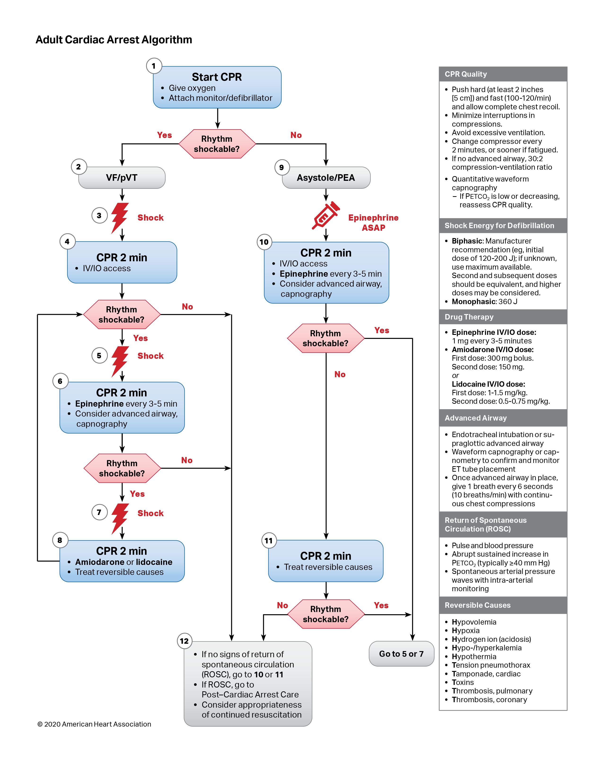
ACLS cardiac arrest algorithm (teaching figure): Use as a visual overview. Energy settings, airway approach, and post-ROSC targets must match your current hospital ACLS card and latest AHA/ILCOR updates — do not rely on this image alone for clinical dosing.
Shockable vs. Non-Shockable Rhythms
Why Shockable Rhythms Matter
Shockable rhythms are abnormal heart rhythms that can be treated with electrical defibrillation. These rhythms have organized electrical activity that can be "reset" by delivering an electric shock, potentially restoring normal sinus rhythm.
Early defibrillation is critical because survival decreases by approximately 10% for every minute that passes without defibrillation in shockable rhythms. The first shock should be delivered within 2 minutes of cardiac arrest recognition.
Defibrillation works by depolarizing all cardiac cells simultaneously, allowing the heart's natural pacemaker (SA node) to regain control and restore organized electrical activity.
Shockable Rhythms
- Ventricular Fibrillation (VF): Chaotic electrical activity, no organized depolarization.
- Pulseless Ventricular Tachycardia (pVT): Organized wide-complex, but no pulse.
- Management: Immediate high-quality CPR → Defibrillation (biphasic energy per manufacturer; often 120–200 J range for first shock — confirm device) → Epinephrine 1 mg IV/IO every 3–5 min → Amiodarone or lidocaine for refractory VF/pVT → continue CPR/defibrillation cycles.
- Post-ROSC care: Support airway/breathing/circulation; treat STEMI urgently; temperature control / avoid fever per current post-arrest guidance and local protocol; hemodynamic optimization and delayed neuroprognostication.
Non-Shockable Rhythms
These rhythms do not respond to electrical defibrillation because they lack organized electrical activity that can be "reset."
- Pulseless Electrical Activity (PEA): Organized electrical activity on monitor, but no mechanical contraction (no pulse).
- Asystole: Flatline - complete absence of electrical activity.
- Management: Continuous CPR → Epinephrine (every 3-5 min) → Focus on the H's and T's (Hypovolemia, Hypoxia, Tamponade, Tension PTX, etc.).
Ventricular Fibrillation (VF)

Chaotic electrical activity with no organized depolarization - shockable rhythm requiring immediate defibrillation to restore normal cardiac rhythm
Asystole
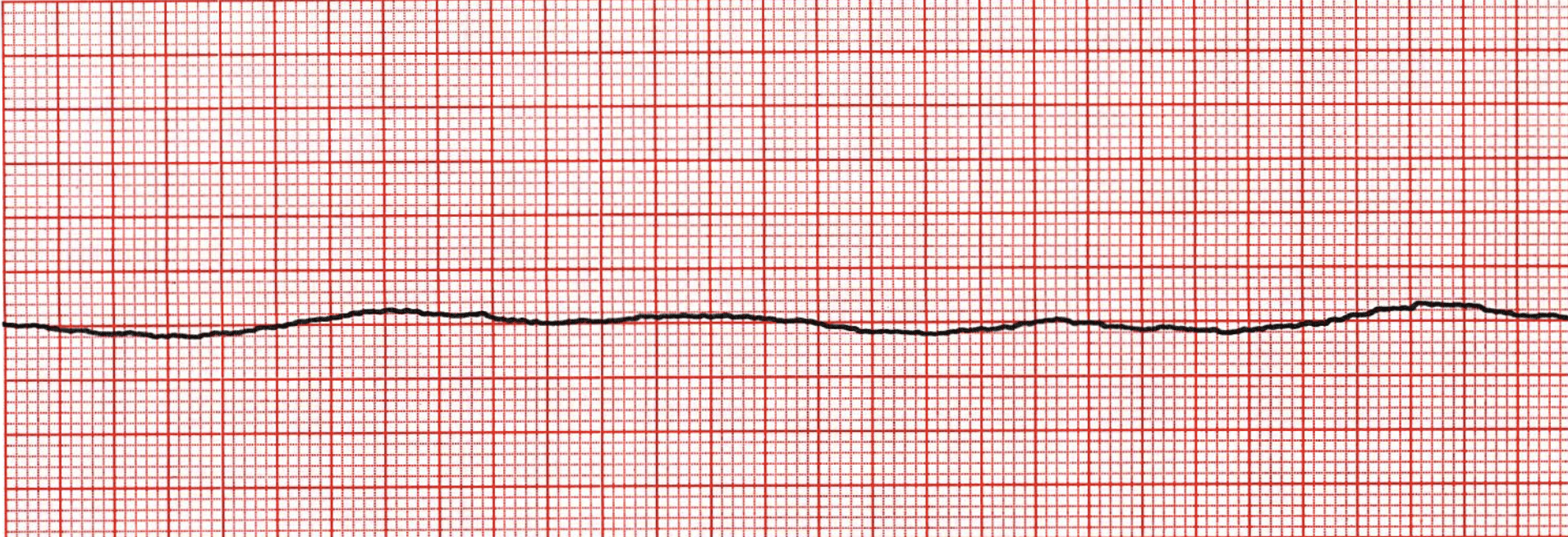
Flatline - No electrical activity visible on ECG monitor
Post-Cardiac Arrest Care
What Happens After ROSC?
The Post-ROSC Algorithm
Time is Brain: The Golden Hour
The first hour after return of spontaneous circulation (ROSC) is critical for neurologic recovery. Immediate optimization of hemodynamics and oxygenation is essential.
0-5 min
Secure airway, optimize oxygenation
5-15 min
12-lead ECG, labs, imaging
15-60 min
Temperature management, cath lab
Immediate Management
Hemodynamic Goals
- Mean BP: ≥ 65 mmHg (MAP)
- SBP: ≥ 90 mmHg
- Urine output: ≥ 0.5 mL/kg/hr
- Lactate clearance as marker
Ventilation Strategy
- ETCO₂: 35-45 mmHg
- SpO₂: 94-98% (avoid hyperoxia)
- Protective ventilation: TV 6-8 mL/kg
- Sedation + analgesia for intubation
Targeted Temperature Management
TTM / temperature control: 32–37.5°C range (protocol-dependent)
- Indicated for: Comatose patients post-OHCA
- Goal: Prevent fever. Target 32–36°C or controlled normothermia <37.5°C (per local protocol / current AHA–ILCOR guidance)
- Duration: Maintain strategy for at least 24h
- Monitor: Shivering, electrolytes, glucose
Neurologic Assessment
- Neuroprognostication: After 72 hours
- SSEP, EEG: For prognostication
- Avoid early withdrawal of care
- Family support throughout
Investigations
Diagnostic Workup
- ECG: STEMI? → Immediate cath lab
- Echo: LV function, tamponade, aortic dissection
- CT chest/abdomen: If indicated
- Coronary angiogram: For OHCA with STEMI
- EEG: For seizure activity
Supportive Care
Medical Management
- Glycemic control: 144-180 mg/dL
- Antiarrhythmics: If recurrent VF/VT
- Antibiotics: If infection suspected
- DVT prophylaxis: When stable
- GI prophylaxis: PPI or H2 blocker
Key Points for Students
- Don't declare brain death early! Wait at least 72 hours post-rewarming
- STEMI post-arrest = cath lab: Even if initially comatose
- Avoid hyperoxia: SpO₂ 94-98% reduces oxidative stress
- Multidisciplinary approach: Cardiology, ICU, neurology involvement
- Family communication: Honest, compassionate, regular updates
ACLS 2: Tachyarrhythmias
Wide vs. Narrow Complex Management
The Unstable Tachycardia Protocol
If the patient is Unstable (hypotension, shock, acute heart failure, ischemic chest pain, altered mental status) due to the rhythm, proceed directly to Synchronized Cardioversion.
Wide Complex Tachycardia (WCT, QRS ≥ 120ms)
- Rule: Treat as Ventricular Tachycardia (VT) until proven otherwise.
- Stable VT: Amiodarone or Procainamide.
- Caution: Avoid AV nodal blockers (Verapamil, Diltiazem) in WCT of unknown origin, especially if WPW is suspected (can precipitate VF).
Narrow Complex Tachycardia (NCT, QRS < 120ms)
- SVT: Stable - Vagal Maneuvers → Adenosine
- A-fib/A-flutter: Stable - Rate Control (Beta-blockers or Calcium Channel Blockers). Check stroke risk (CHADS2-VASc).
Ventricular Tachycardia (VT)
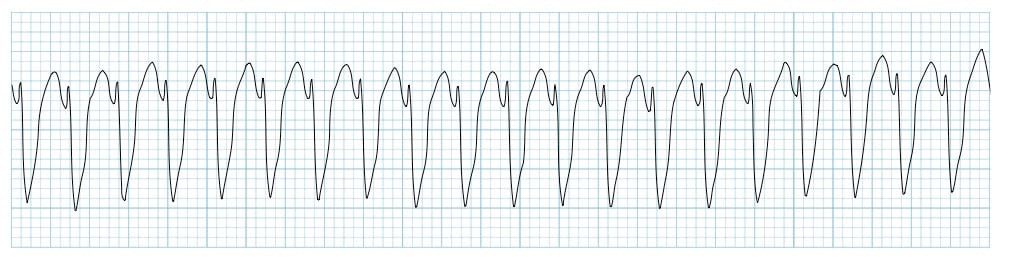
Wide complex tachycardia (QRS ≥ 120ms) with regular rhythm - treat as VT until proven otherwise
Atrial Fibrillation in the ED
Rapid Diagnosis & Management
AF Recognition & Initial Assessment
Clinical Presentation
- Irregularly irregular pulse - hallmark finding
- Palpitations - most common complaint
- Dyspnea, chest pain, fatigue
- May be asymptomatic - discovered incidentally
ECG Features
- Irregular R-R intervals
- Absence of P waves
- Coarse or fine atrial fibrillation waves
- Variable ventricular response
Critical Initial Steps
1. ABCs
Assess stability - rate-related symptoms?
2. Duration
< 48h vs > 48h (anticoagulation implications)
3. Stroke Risk
CHA₂DS₂-VASc score
Rate vs Rhythm Control
Rate Control (Preferred for ED)
- β-blockers: Metoprolol 2.5-5mg IV, or PO
- CCBs: Diltiazem 10-20mg IV (avoid in HF)
- Digoxin: 0.25mg IV (renal/hepatic caution)
- Goal: HR < 110 bpm at rest
Rhythm Control (Selected cases)
- Stable patient with recent onset
- Amiodarone: 150mg IV over 10 min
- Cardioversion: if unstable
Anticoagulation Strategy
CHA₂DS₂-VASc Score ≥2
- New onset AF: Start anticoagulation
- DOAC preferred: Apixaban or Rivaroxaban
- Warfarin: If CrCl <30 or mechanical valve
AF Duration <48h
- Low stroke risk: Can cardiovert without AC
- Start AC: After cardioversion
- TEE: If duration uncertain
Clinical Pearls for Students
- Don't miss the irregular pulse! Feel the radial pulse for 30 seconds
- Rate control first: Most ED patients don't need rhythm control
- Stroke prevention: Address anticoagulation even if rhythm restored
- Follow-up: All patients need cardiology referral
ACLS 3: Bradyarrhythmias & Blocks
When to Pace, When to Wait
Symptomatic Bradycardia Algorithm
The Need for Intervention
- Bradycardia requires treatment only if it is causing symptoms: Shock/Hypotension, Acute Altered Mental Status, Acute Heart Failure, or Ischemic Chest Pain.
- High-Grade Blocks: Mobitz Type II and 3rd Degree Heart Block are inherently unstable and often require pacing.
Management Steps
- Step 1 (First Line): Atropine (1mg IV, max 3mg). Less effective for infra-nodal blocks (Type II/3rd Degree).
- Step 2 (If Atropine fails): Transcutaneous Pacing (TCP). Start at 60-80 bpm, increase mA until capture.
- Step 3 (Alternatives/Bridge): Dopamine or Epinephrine infusion (chronotropic agents).
Bradycardia ECG Patterns
Recognition and Management of Bradycardic Rhythms
Common Bradycardia ECG Patterns
Bradycardia ECG Patterns
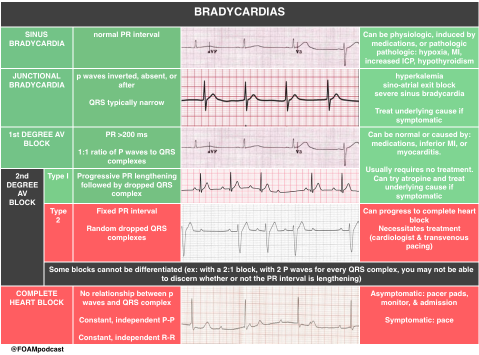
Common Bradycardia Patterns:
Sinus Bradycardia
- • Normal P waves before each QRS
- • Rate < 60 bpm
- • Often benign if asymptomatic
Heart Block
- • P waves and QRS dissociated
- • May be 2nd or 3rd degree
- • Requires pacing if symptomatic
⚠️ Bradycardia requires treatment only if symptomatic. Immediate pacing may be needed for high-grade blocks or hemodynamic instability.
Heart Failure
Acute Decompensated Heart Failure (ADHF)
💔 Heart Failure: When the Pump Fails
What is Heart Failure?
Heart failure is a clinical syndrome characterized by the heart's inability to pump blood effectively to meet the body's metabolic demands. It can be acute or chronic, systolic or diastolic.
Systolic HF
Reduced ejection fraction (<40%). "Weak pump" - impaired contraction.
Diastolic HF
Preserved ejection fraction (≥50%). "Stiff pump" - impaired relaxation/filling.
Clinical Presentation
Cardinal Symptoms
- 🫁 Dyspnea: Orthopnea, PND (paroxysmal nocturnal dyspnea)
- 💧 Peripheral edema: Sacral, pedal edema
- 😮 Fatigue: Reduced exercise tolerance
- 💓 Tachycardia: Compensatory response
Respiratory Signs
- 🫁 Pulmonary rales: Crackles on auscultation
- 💨 S3 gallop: Low-frequency extra heart sound
- 🫀 Jugular venous distension: Elevated JVP
- 📏 Hepatojugular reflux: Positive test
NYHA Classification
Management Principles
- 💧 Diuretics: Loop diuretics (furosemide) for congestion
- 💊 Vasodilators: Nitroglycerin for preload reduction
- 🫀 Inotropes: Dobutamine for severe cases
- 📊 Monitor: Daily weights, fluid balance
Red Flags: Immediate Intervention
- • Hypotension: SBP <90 mmHg with symptoms
- • Acute pulmonary edema: Respiratory distress
- • Cardiorenal syndrome: Rising creatinine
- • Arrhythmias: New-onset atrial fibrillation
- • Progression to cardiogenic shock
Cardiogenic Shock
The Failing Pump: Emergency Management
🚨 Cardiogenic Shock: Pump Failure with Systemic Impact
What is Cardiogenic Shock?
Cardiogenic shock is a state of critical end-organ hypoperfusion due to primary cardiac dysfunction. It's characterized by inadequate tissue perfusion despite adequate preload, resulting in cellular hypoxia and organ dysfunction.
Clinical Features
Hemodynamic Criteria
- 📊 CI <2.2 L/min/m²: Low cardiac index
- 💉 MAP <65 mmHg: Mean arterial pressure
- 🫀 PCWP >15 mmHg: Elevated wedge pressure
- ⚡ SVR normal or ↑: Systemic vascular resistance
End-Organ Effects
- 🧠 Confusion/altered mental status
- 💛 Oliguria (<0.5 mL/kg/hr)
- 🫁 Pulmonary congestion/edema
- 🌡️ Cool extremities, delayed capillary refill
Causes of Cardiogenic Shock
Emergency Management
1. ABCs + Oxygen
2. Identify & treat reversible causes
3. Inotropic support: Dobutamine ± norepinephrine
4. Mechanical support: IABP, Impella, ECMO
5. Definitive therapy: PCI, CABG, valve repair
Mortality: 40-50% Despite Treatment
- • Early recognition is critical - intervene before end-organ damage
- • SCAI classification: A (at risk) → B (beginning) → C (classic) → D (deteriorating) → E (extremis)
- • Time to intervention: Door-to-support time should be <90 minutes
- • Multidisciplinary approach: Cardiology, cardiac surgery, critical care
Obstructive Cardiac Emergencies
Tamponade, Pulmonary Embolism & Mechanical Obstruction
🔒 Obstructive Shock: When the Pump Can't Fill or Empty
Cardiac Tamponade
Beck's Triad
- 🔽 Hypotension: Low blood pressure
- 🫁 JVD: Jugular venous distension
- ❤️ Muffled heart sounds: Distant, quiet
Pulsus Paradoxus
>10 mmHg drop in SBP during inspiration
POCUS Findings
RV diastolic collapse, swinging heart
Tamponade ECHO
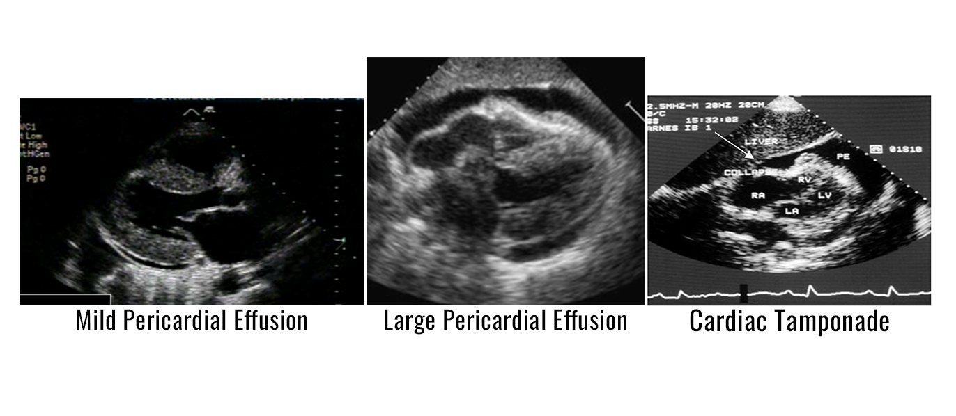
Pericardial effusion causing RV collapse
Emergency Management
- 🔴 Immediate: IV fluids + vasopressors
- 🟡 Definitive: Pericardiocentesis (ECHO-guided)
- 🟢 Avoid: Positive pressure ventilation
Pulmonary Embolism (PE)
Massive PE Criteria
- 🔽 SBP <90 mmHg for ≥15 min
- 🫁 Respiratory failure (O2 sat <90%)
- 💙 RV dysfunction on ECHO
- 🏥 ICU admission required
Time is Lung
Door-to-needle:
<15 min for massive PE
Thrombolytics: tPA 100mg IV
over
2h
Clinical Clues
- • Sudden dyspnea, chest pain
- • Tachycardia, hypoxia
- • Unilateral leg swelling
- • Risk factors: Immobility, cancer, OCP
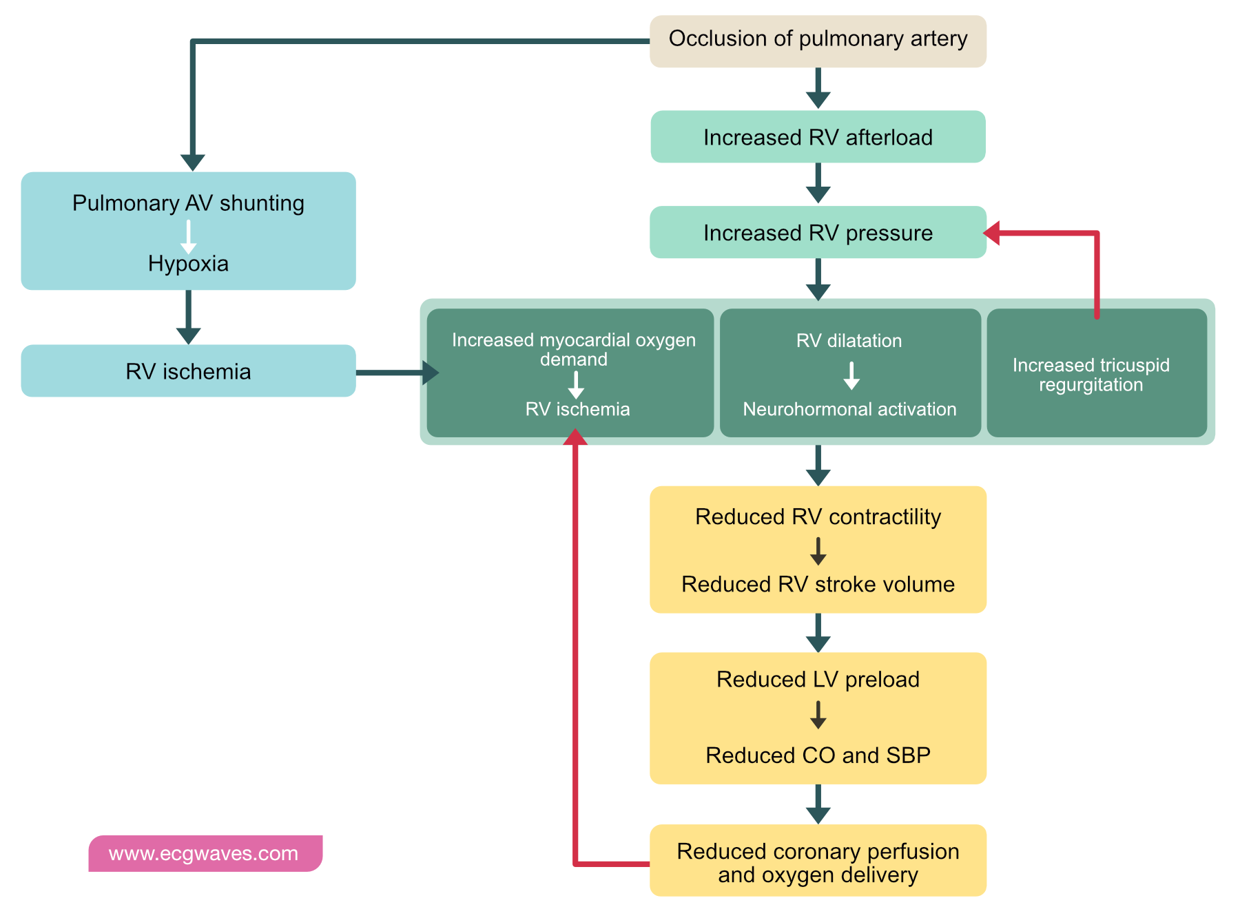
PE Pathophysiology
Clot obstructs pulmonary artery → RV strain → shock
Management Algorithm
1. ABCs + Oxygen
2. Assess stability:
• Stable: Anticoagulation
• Unstable: Thrombolytics
3. Consider thrombectomy if contraindications
Acute Pericarditis
Inflammation of the Pericardial Sac
🔥 Pericarditis: When the Heart's Wrapper is on Fire
Pericarditis Diagnostic Checklist
Diagnosis: ≥2 of 4 criteria + typical chest pain. ECG changes are highly specific!
Clinical Presentation
Chest Pain Characteristics
- 🔥 Sharp, pleuritic (worse with inspiration)
- 📍 Retrosternal or left precordial
- 🏃♂️ Better with sitting/leaning forward
- ⏰ Can last hours to days
Physical Exam
- 🔊 Pericardial friction rub: "Scratchy" sound
- 🌡️ Fever in infectious cases
- 🫁 Tachypnea if pleural effusion
- 💓 Distant heart sounds if effusion
Classic Pericarditis ECG
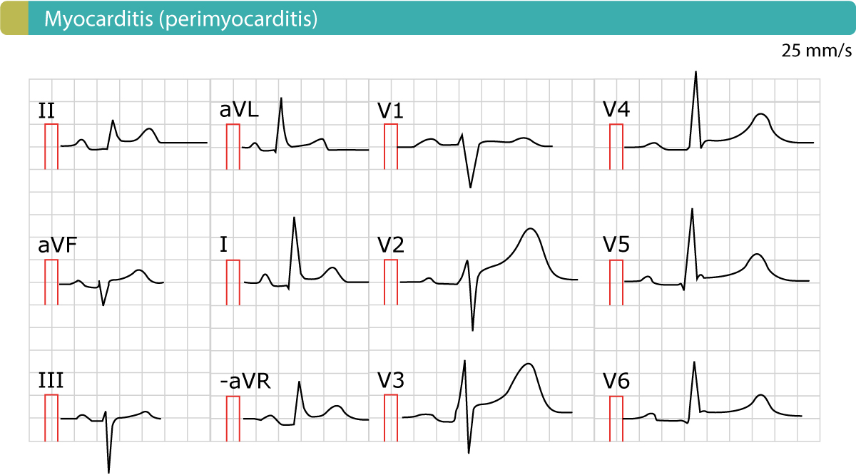
Diffuse ST elevation + PR depression
Treatment Protocol
1st Line: NSAIDs (Ibuprofen 600-800mg TID) + PPI
Add-on: Colchicine 0.5mg BID (reduces recurrence)
Steroids: Only if contraindications to NSAIDs
Duration: 1-2 weeks, taper gradually
Red Flags - Seek Immediate Care
- • Tamponade signs: Hypotension, JVD, pulsus paradoxus
- • Large effusion: >2cm on ECHO
- • Myocarditis: Elevated troponins, LV dysfunction
- • Recurrent episodes: Multiple flares
Acute Myocarditis
Inflammation of the Heart Muscle
💔 Myocarditis: When the Heart Muscle is Under Attack
Myocarditis Risk Stratifier
Low Risk
- • Normal EF (>50%)
- • No arrhythmias
- • Mild symptoms
- • Troponin ≤3x URL
Outpatient management
Intermediate Risk
- • EF 35-50%
- • Non-sustained VT
- • Moderate symptoms
- • Troponin 3-10x URL
Telemetry monitoring
High Risk
- • EF <35%
- • Sustained VT/VF
- • Cardiogenic shock
- • Troponin >10x URL
ICU admission
Causes & Presentation
Common Causes
- 🦠 Viral: Coxsackie, EBV, CMV (most common)
- 💊 Drugs: Doxorubicin, cocaine, NSAIDs
- 🦠 Bacterial: Lyme, diphtheria, sepsis
- ⚡ Autoimmune: SLE, sarcoidosis
- 🔬 Hypersensitivity: Sulfa drugs, vaccines
Clinical Signs
- 💔 Chest pain: Retrosternal, worse supine
- 😮 Dyspnea: Acute heart failure
- 💓 Palpitations: Arrhythmias common
- 🌡️ Fever: Infectious cases
- ⚡ Fatigue: Viral prodrome
Myocarditis ECG Changes
ST changes, conduction blocks, arrhythmias
Management Strategy
🔴 Supportive Care: Rest, monitor hemodynamics
🟡 Heart Failure: ACEi/ARBs, beta-blockers, diuretics
🟢 Arrhythmias: Antiarrhythmics as needed
🔵 MCS: IABP/ECMO for refractory shock
🟣 Transplant: Bridge to recovery/transplant
Diagnostic Workup
Essential Tests:
- • Troponin (elevated in 90%)
- • BNP/NT-proBNP
- • ECG (daily monitoring)
- • ECHO (EF assessment)
Advanced Testing:
- • Cardiac MRI (gold standard)
- • Endomyocardial biopsy
- • Viral PCR studies
- • Autoimmune workup
Valvular Heart Emergencies
Acute Valve Dysfunction in the ED
🚪 Valvular Emergencies: When Heart Valves Fail Suddenly
Decompensated Aortic Stenosis
Emergency Presentation
- 💔 Acute heart failure: Pulmonary edema
- 🔽 Hypotension: Fixed cardiac output
- 🫁 Dyspnea: LV failure symptoms
- 💓 Angina: Supply-demand mismatch
Classic Physical Exam
- 🔊 Harsh systolic murmur: RUSB/base
- 📉 Narrow pulse pressure: Fixed stroke volume
- 💓 Slow rising carotid pulse: "Parvus et tardus"
- 🫀 LV heave: Pressure overload
Management
- 🔴 Stabilize: Vasopressors for hypotension
- 🟡 Avoid: Nitroglycerin, beta-blockers
- 🟢 Definitive: Urgent surgical consultation
- 🔵 TAVR: Alternative for high-risk patients
Acute Mitral Regurgitation
Causes
- 🔗 Chordae rupture: Endocarditis, trauma
- 💔 Papillary muscle rupture: MI complication
- ⚡ Acute ischemia: papillary muscle dysfunction
- 🔄 Prosthetic valve dysfunction
Presentation
- 🫁 Acute pulmonary edema: Volume overload
- 💓 Holosystolic murmur: Apex, radiates axilla
- 🔊 S3 gallop: Rapid filling
- 📉 Hypotension: Low cardiac output
Emergency Management
- 🔴 Vasopressors: Maintain BP (afterload)
- 🟡 IABP: Reduces afterload temporarily
- 🟢 Definitive: Urgent surgical repair
- 🔵 Avoid: Excessive vasodilation
Prosthetic Valve Emergencies
Thrombosis
- ❌ INR <2.5: Mechanical valves
- 🔊 New murmur: Regurgitation
- 🫁 Pulmonary edema: Acute failure
- 🔴 Embolic events: Stroke risk
Endocarditis
- 🌡️ Fever: Persistent bacteremia
- 💓 New murmur: Perivalvular leak
- 🔴 Emboli: Septic emboli
- 🫁 Abscess: Periannular complications
Structural Failure
- 🔊 Clicking sound: Leaflet fracture
- 💓 Regurgitant murmur: Paravalvular leak
- 🫁 Hemolysis: Anemia, jaundice
- 📉 Cardiac failure: Acute decompensation
Management
Thrombosis: Heparin + urgent surgery
Endocarditis: Broad-spectrum ABX + surgery
Structural: TEE-guided intervention
All cases: Urgent cardiac surgery consult
Aortic Emergencies
Dissection and Ruptured Abdominal Aortic Aneurysm (AAA)
The Vascular Catastrophes
Aortic Dissection
- Pain: Sudden, severe, tearing/ripping chest/back pain.
- Exam: Look for Pulse/BP Differential (>20 mmHg difference between arms) and new Aortic Regurgitation murmur.
- Tx: Aggressive Heart Rate (≤ 60 bpm) and Blood Pressure (SBP ≤ 120 mmHg) control (Esmolol, Labetalol). Emergent Type A surgery.
Ruptured AAA
- Triad: Pain (abdominal/flank/back), Pulsatile abdominal mass, and Hypotension. (Only present in ~ 50% of cases).
- Key: Consider in all elderly patients with back/abdominal pain and hypotension/syncope.
- Tx: Immediate surgical consult. Permissive Hypotension (target SBP ~ 90 mmHg) to avoid dislodging clot.
Hypertensive Emergencies
Urgency vs. Emergency
Diagnosis and Treatment Goals
Hypertensive Emergency
- Definition: Severe hypertension (≥ 180/120 mmHg) PLUS acute end-organ damage (e.g., encephalopathy, stroke, acute HF, ACS, aortic dissection, acute renal failure).
- Goal: Reduce MAP by 10-15% in the first hour, and by max 25% in the first day. (Exceptions: Aortic Dissection requires *immediate* SBP ≤ 120 mmHg).
- Tx: IV titratable agents (Labetalol, Nicardipine, Esmolol).
Hypertensive Urgency
- Definition: Severe hypertension (≥ 180/120 mmHg) WITHOUT evidence of acute end-organ damage.
- Goal: Do NOT acutely lower BP in the ED. Risk of cerebral hypoperfusion outweighs benefits.
- Tx: Restart home meds or start oral agent. Discharge with close outpatient follow-up.
Syncope 1: Etiologies & Risk
Transient Loss of Consciousness (T-LOC)
Three Major Categories of Syncope
Reflex (Neurally Mediated)
- Vasovagal: Classic faint, triggered by stress, pain, or sight of blood. Prodrome (nausea, diaphoresis).
- Situational: Triggered by cough, micturition, defecation.
- Carotid Sinus: Triggered by neck compression/shaving.
Orthostatic Hypotension
- Caused by failure of baroreflex → hypotension upon standing.
- Etiologies: Volume depletion (dehydration), medications (diuretics, antihypertensives), autonomic dysfunction (DM, Parkinson's).
- Diagnosis: Drop of SBP ≥ 20 or DBP ≥ 10 mmHg within 3 minutes of standing.
Cardiac Syncope (High Risk)
- Arrhythmic: VT, high-grade blocks. Often abrupt, without prodrome.
- Structural: Severe Aortic Stenosis, HOCM, Tamponade, Pulmonary Embolism (obstructive).
- Key: Cardiac syncope is ~ 5 times more likely to result in adverse events than non-cardiac.
Syncope 2: The Evaluation
Finding the High-Risk Patient
Red Flags & High-Risk Features
Cardiac Red Flags
- Syncope during exertion (structural heart disease, increased risk).
- Syncope while supine (arrhythmia, increased risk).
- Associated symptoms: Chest pain, dyspnea, palpitations prior to LOC.
- Family history of sudden cardiac death.
Essential Initial Workup
- ECG: Must rule out blocks, VT/VF history, long QT, or WPW.
- Orthostatic Vitals: Must be performed in all patients.
- Labs: Hgb (anemia), Troponin (ischemia), B-HCG (pregnancy).
- POCUS: If heart failure or structural disease is suspected.
Canadian Syncope Risk Score (CSRS)
A validated clinical decision tool to identify patients at low risk for serious outcomes within 30 days:
Low Risk (Score -3 to 0):
- • No serious underlying diagnosis
- • No adverse events at 30 days
- • Safe for discharge with follow-up
High Risk (Score ≥1):
- • 2.5% risk of serious outcome
- • Requires hospital admission
- • Further cardiac evaluation needed
Disposition Decision
Hospital admission is required for all patients with cardiac syncope or those with high-risk features (e.g., HF history, abnormal ECG, exertional syncope), even if the initial workup is non-diagnostic.
Cardiac Medications in the ED
Common Drugs & Their Indications
Essential Cardiac Drugs for Emergency Use
Vasopressors & Inotropes
Norepinephrine (Levophed)
- Indication: Hypotension, septic/cardiogenic shock
- Dose: 0.05-0.5 mcg/kg/min IV
- Effects: α₁ > β₁ (vasoconstriction)
- Caution: Monitor extremities for ischemia
Epinephrine
- ACLS arrest: 1 mg IV/IO every 3–5 min (all arrest rhythms)
- Anaphylaxis (adult IM): 0.3–0.5 mg IM (1:1000) — different concentration/route than arrest dosing
- β₁β₂α₁ effects: Inotropy + chronotropy + vasoconstriction (dose-dependent)
- Infusion: titrate (commonly started ~0.05–0.5 mcg/kg/min) per protocol
Dopamine
- Second-line agent: After Norepinephrine
- Medium (5-10 mcg/kg/min): Inotropy (β₁)
- High (10-20 mcg/kg/min): Vasoconstriction (α₁)
- Note: NO "Renal Dose" benefit
Antiarrhythmics
Amiodarone
- Refractory VF/pVT: 300 mg IV/IO first, then 150 mg
- Stable wide-complex / AF (selected): often 150 mg IV over 10 min — indication-specific
- Multi-channel blocker (Class III predominant teaching label)
- Caution: Hypotension, bradycardia, long half-life
Lidocaine
- Refractory VF/pVT option: 1–1.5 mg/kg IV/IO, then 0.5–0.75 mg/kg
- Class Ib agent (Na⁺ channel)
- Alternative/adjunct option to amiodarone in ACLS algorithms
- Caution: CNS toxicity at high cumulative doses
Adenosine
- Regular narrow-complex SVT: 6 mg rapid IV push, then 12 mg (if needed)
- Half-life: <10 seconds
- Diagnostic for SVT vs VT
- Caution: WPW (can cause VF)
Antihypertensives
Labetalol
- HTN emergency: 20mg IV bolus
- Aortic dissection: Preferred agent
- α₁β₁β₂ blocker
- Caution: Asthma, bradycardia
Nicardipine
- HTN emergency: 5mg/hr IV infusion
- CCB - pure vasodilator
- Less reflex tachycardia
- Titratable, predictable
Anticoagulants & Antiplatelets
Heparin
- STEMI/NSTEMI: 60 IU/kg IV bolus
- LMWH alternative: Enoxaparin
- Monitor: aPTT 1.5-2x normal
- Reversal: Protamine
Antiplatelets
- Aspirin: 325mg PO/NG chewed
- Clopidogrel: 300-600mg loading
- Ticagrelor: 180mg loading
- Give before cath when possible
Medication Mnemonics for Students
Pressor Order
"LED" = Levophed → Epinephrine → Dopamine
(Increasing vasoconstriction)
AF Rate Control
"BBD" = Beta-blockers → CCBs → Digoxin
(Decreasing renal function)
Antiarrhythmics
"A Dozen Little Children" = Amiodarone, Dofetilide, Lidocaine, etc.
HTN Emergency
"LIP" = Labetalol, Nicardipine, Nitroprusside
Critical Pearls
- Know your doses cold! Practice ACLS algorithms regularly
- Double-check calculations - medication errors kill
- Monitor for side effects: Hypotension, bradycardia, torsades
- Reversal agents: Know common reversal agents for anticoagulants you use
- Drug interactions: Consider comorbidities and other meds
Summary
Key Takeaways from Critical Cardiac Emergencies
Essential Points to Remember
Time-Critical Interventions
- Door-to-device (STEMI): system goal often ≤90 min (transfer pathway often ≤120 min FMC-to-device)
- Door-to-needle: ≤30 min when fibrinolysis is the chosen strategy
- Defibrillation: biphasic energy per manufacturer (often ~120–200 J first shock); minimize delay
- Post-ROSC: prevent fever / TTM per protocol (e.g. 32–36°C or <37.5°C), early PCI for STEMI-equivalent
- Early recognition saves lives
Critical Assessments
- ECG within 10 minutes for high-risk presentations
- Beck's Triad for tamponade
- Orthostatic vitals for all syncope patients
- Canadian Syncope Risk Score for disposition decisions
- Serial reassessment - conditions evolve
Essential Medications
- AF Rate Control: β-blockers first-line
- STEMI Cocktail: Aspirin + Heparin + Antiplatelets
- Shock: Norepinephrine for hypotension
- VT/VF: Amiodarone or Lidocaine
- Know your doses! Practice prevents errors
Remember the H's and T's
This comprehensive presentation covers the complete spectrum of critical cardiac emergencies from ACS to arrhythmias, tamponade to aortic dissection, with special focus on ED-specific management and the latest evidence-based protocols.
New topics added: Atrial Fibrillation in the ED • Post-Cardiac Arrest Care • Cardiac Medications • Code STEMI Activation
Always consult local protocols and consider patient-specific factors in clinical decision-making.
Thank You!
Critical Cardiac Emergencies Presentation Complete
Thank you for your attention and dedication to saving lives!
You've completed the comprehensive guide to Critical Cardiac Emergencies. Remember: Every second counts in cardiac emergencies - your knowledge can make the difference between life and death.
Selected References
- AHA CPR & ECC Guidelines (ACLS / resuscitation)
- ACC/AHA Chest Pain & ACS guidance (incl. 2021 chest pain)
- ESC 2023 Acute Coronary Syndromes Guidelines
- LITFL ECG Library
- GrepMed Clinical Visuals
Educational resource only (SASEM). Algorithms and doses can change — always verify against the latest AHA/ESC/ACC publications and your hospital protocol. Content reviewed for structure 2026; re-check focused ACLS updates regularly.
Follow on X (Twitter)
@I_am_Doctor